
File:Histology bse.jpg
此为最大尺寸。
Histology_bse.jpg (700 × 558像素,文件大小:73 KB,MIME类型:image/jpeg)
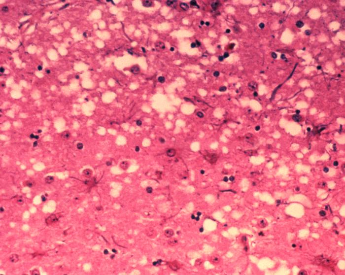
| 描述 |
English: This micrograph of brain tissue reveals the cytoarchitectural histopathologic changes found in bovine spongiform encephalopathy. The presence of vacuoles, i.e. microscopic “holes” in the gray matter, gives the brain of BSE-affected cows a sponge-like appearance when tissue sections are examined in the lab.
Nederlands: Deze microscopische opname toont hersenweefsel van een koe die aan BSE gestorven is. Tussen de hersencellen ziet men duidelijk verschillende vacuoles, die deze coupe (weefselsnede) een sponsachtig aanzicht geven.
Deutsch: Das Bild zeigt die histopathologischen Veränderungen die bei einer Infektion mit BSE auftreten. Die Vakuolen, die in der grauen Substanz (substantia grisea) auftreten geben dem Bild ein schwamm-artiges Aussehen.
Français : Cette coupe de tissu cérébral montre les modifications histopathologiques de l'organisation cellulaire lors d'une encéphalopathie spongiforme bovine. la présence de vacuoles, c'est-à-dire des "trous" microscopiques dans le tissu cérébral, donne au cerveau de vaches atteintes de l'ESB un aspect en éponge à l'examen des tissus en laboratoire. |
| 日期 | |
| 来源 | Public Health Image Library, APHIS: http://www.aphis.usda.gov/lpa/issues/bse/bse_photogallery.html |
| 作者 | Dr. Al Jenny |
| 其他版本 | http://en.wikipedia.org/wiki/Image:Aphis.usda.gov_BSE_5.jpg |
|
|
 |
文件历史
点击某个日期/时间查看对应时刻的文件。
| 日期/时间 | 缩略图 | 大小 | 用户 | 备注 | |
|---|---|---|---|---|---|
| 当前 | 2005年6月21日 (二) 10:21 | | 700 × 558(73 KB) | Obarskyr | {{PD}} |
文件用途
以下页面使用本文件:
全域文件用途
以下其他wiki使用此文件:
- af.wikipedia.org上的用途
- ast.wikipedia.org上的用途
- as.wikipedia.org上的用途
- bn.wikipedia.org上的用途
- bs.wikipedia.org上的用途
- cs.wikipedia.org上的用途
- de.wikipedia.org上的用途
- de.wikibooks.org上的用途
- el.wikipedia.org上的用途
- en.wikipedia.org上的用途
- en.wikibooks.org上的用途
- en.wikinews.org上的用途
- eo.wikipedia.org上的用途
- es.wikipedia.org上的用途
- eu.wikipedia.org上的用途
- fa.wikipedia.org上的用途
- fi.wiktionary.org上的用途
- fr.wikipedia.org上的用途
- gl.wikipedia.org上的用途
- he.wikipedia.org上的用途
- ht.wikipedia.org上的用途
- hy.wikipedia.org上的用途
- ia.wikipedia.org上的用途
- id.wikipedia.org上的用途
- is.wikipedia.org上的用途
- it.wikipedia.org上的用途
- ja.wikipedia.org上的用途
- jv.wikipedia.org上的用途
- ko.wikipedia.org上的用途
- lt.wikipedia.org上的用途
- ms.wikipedia.org上的用途
- nl.wikipedia.org上的用途
- no.wikipedia.org上的用途
- oc.wikipedia.org上的用途
- pl.wikipedia.org上的用途
- pnb.wikipedia.org上的用途
查看本文件的更多全域用途。