
File:Validation of the dye diffusion assay performed with the flattened cochlear preparation.png

本预览的尺寸:287 × 599像素。 其他分辨率:115 × 240像素 | 230 × 480像素 | 979 × 2,044像素。
原始文件 (979 × 2,044像素,文件大小:3.33 MB,MIME类型:image/png)
摘要
| 描述 |
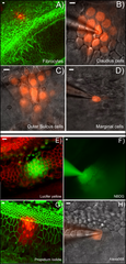
English: A–D: Dye diffusion patterns after PI was injected into a single cell in various locations in the cochlea. The type of the cells that was injected is given at lower right corner of each panel. E–F: Diffusion patterns of four different fluorescent dyes after injecting into a single Claudius cell. Name of the dye is given in the lower right corner of each panel. Panels B), C), D), F) & H) were photographed with unfixed fresh samples. Panels A), E), G) were results obtained from fixed samples after the experiments were done. They were labeled with fluorescent phalloidin (red in E, green in A&G) to outline the cell border. Scale bar on the top left of each panel represents approximately 100 µm. |
| 日期 | |
| 来源 | PLOS ONE an open source peer reviewed journal- Gap Junction Mediated Intercellular Metabolite Transfer in the Cochlea Is Compromised in Connexin30 Null Mice ([1]) |
| 作者 | Qing Chang, Wenxue Tang, Shoeb Ahmad1, Binfei Zhou1, Xi Lin1 |
许可协议
文件历史
点击某个日期/时间查看对应时刻的文件。
| 日期/时间 | 缩略图 | 大小 | 用户 | 备注 | |
|---|---|---|---|---|---|
| 当前 | 2009年2月12日 (四) 04:24 | | 979 × 2,044(3.33 MB) | Mike.lifeguard | {{Information |Description={{en|1=A–D: Dye diffusion patterns after PI was injected into a single cell in various locations in the cochlea. The type of the cells that was injected is given at lower right corner of each panel. E–F: Diffusion patterns o |
文件用途
以下页面使用本文件:
全域文件用途
以下其他wiki使用此文件:
- en.wikipedia.org上的用途
- pt.wikipedia.org上的用途